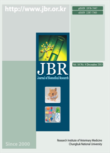
간행물
Journal of Biomedical Research

- 발행기관 충북대학교 동물의학연구소
- 자료유형 학술지
- 간기 계간
- ISSN 1976-7447 (Print)2287-7363 (Online)
- 수록기간 2000 ~ 2015
- 주제분류 의약학 > 수의학 의약학 분류의 다른 간행물
- 십진분류KDC 528DDC 636
권호리스트/논문검색
Vol. 16 No. 3 (2015년 9월) 9건
Review
1.
2015.09
구독 인증기관 무료, 개인회원 유료
Speech and language are uniquely human-specific traits that have contributed to humans becoming the predominant species on earth from an evolutionary perspective. Disruptions in human speech and language function may result in diverse disorders, including stuttering, aphasia, articulation disorder, spasmodic dysphonia, verbal dyspraxia, dyslexia, and specific language impairment (SLI). These disorders often cluster within a family, and this clustering strongly supports the hypothesis that genes are involved in human speech and language functions. For several decades, multiple genetic studies, including linkage analysis and genome-wide association studies, were performed in an effort to link a causative gene to each of these disorders, and several genetic studies revealed associations between mutations in specific genes and disorders such as stuttering, verbal dyspraxia, and SLI. One notable genetic discovery came from studies on stuttering in consanguineous Pakistani families; these studies suggested that mutations in lysosomal enzyme-targeting pathway genes (GNPTAB, GNPTG, and NAPGA) are associated with non-syndromic persistent stuttering. Another successful study identified FOXP2 in a Caucasian family affected by verbal dyspraxia. Furthermore, an abnormal ultrasonic vocalization pattern (USV) was observed in knock-in (KI) and humanized mouse models carrying mutations in the FOXP2 gene. Although studies have increased our understanding of the genetic causes of speech and language disorders, these genes can only explain a small fraction of all disorders in patients. In this paper, we summarize recent advances and future challenges in an effort to reveal the genetic causes of speech and language disorders in animal models.
4,000원
Original Article
2.
2015.09
구독 인증기관 무료, 개인회원 유료
Vikash Kumar Shah, Sam-Shik Na, Myong-Soo Chong, Jae-Hoon Woo, Yeong-Ok Kwon, Mi Kyeong Lee, Ki-Wan Oh
Poria cocos is a well-known traditional Chinese traditional medicine (TCM) that grows around roots of pine trees in China, Korea, Japan, and North America. Poria cocos has been used in Asian countries to treat insomnia as either a single herb or part of an herbal formula. In a previous experiment, pachymic acid (PA), an active constituent of Poria cocos ethanol extract (PCE), increased pentobarbital-induced sleeping behaviors. The aim of this experiment was to evaluate whether or not PCE and PA modulate sleep architectures in rats as well as whether or not their effects are mediated through GABAA-ergic transmission. PCE and PA were orally administered to individual rats 7 days after surgical implantation of a transmitter, and sleep architectures were recorded by Telemetric Cortical encephalogram (EEG) upon oral administration of test drugs. PCE and PA increased total sleep time and non-rapid eye movement (NREM) sleep as well as reduced numbers of sleep/wake cycles recorded by EEG. Furthermore, PCE increased intracellular chloride levels, GAD65/67 protein levels, and α-, β-, and γ-subunits of GABAA receptors in primary cultured hypothalamic neuronal cells. These data suggest that PCE modulates sleep architectures via activation of GABAA-ergic systems. Further, as PA is an active component of PCE, they may have the same pharmacological effects.
4,000원
3.
2015.09
구독 인증기관 무료, 개인회원 유료
Regarding therapies for treatment of corneal wounds, ex vivo corneal culture is the most effective for minimizing expensive animal studies. Eighteen porcine enucleated eyes were soaked in 0.2% povidone iodine solution for disinfection prior to cornea excision. Subsequently, corneas were excised from whole eyes and filled with an agar/medium mixture. Corneas were transferred into culture dishes, after which culture medium was added until the limbus was covered. Cultures were then placed onto a plate rocker to mimic blinking action, followed by incubation at 37°C and 5% CO2. Corneas were harvested on Days 0, 3, and 7 after incubation, and optical coherence tomography (OCT) was performed on Day 7. Two eyes from each group were fixed in 2% glutaraldehyde/4% paraformaldehyde for low-vacuum scanning electron microscopy (LV-SEM), and four eyes from each group were fixed in 10% neutral-buffered formalin for histological analysis. OCT results showed that central corneal thickness significantly increased by Day 7 compared to Day 0 (P<0.05). Using LV-SEM, gaps between endothelial cells were detected on Day 7 of ex vivo culture. In the histological evaluation, four to five stratified squamous cell layers, wing cells, and basal cells in the epithelium as well as flat-shaped keratocytes in the stroma were found on Day 0. By Day 7, stratified squamous cells and basal cells had decreased in number, and slightly round-shaped keratocytes were observed; however, the number of keratocytes was similar to that on Day 0. In this short-term ex vivo culture, epithelium and endothelium were sensitive to culture, whereas stroma and keratocytes were well maintained. An additional deswelling method will be needed to obtain more successful results in porcine corneal ex vivo culture.
4,000원
4.
2015.09
구독 인증기관 무료, 개인회원 유료
Tight junctions (TJs) form continuous intercellular contacts in intercellular junctions. TJs involve integral proteins such as occludin (OCLN) and claudins (CLDNs) as well as peripheral proteins such as zona occludens-1 (ZO-1) and junctional adhesion molecules (JAMs). TJs control paracellular transportation across cell-to-cell junctions. Although TJs have been studied for several decades, comparison of the transcriptional-translational levels of these molecules in canine organs has not yet been performed. In this study, we examined uterine expression of CLDNs, OCLN, junction adhesion molecule-A, and ZO-1 in canine. Expression levels of canine uterine TJ proteins, including CLDN1, 2, 4, 5, JAM-A, ZO-1, and OCLN, were measured using reverse transcription PCR, real-time PCR, and Western blotting, whereas TJs distribution was determined by immunohistochemistry. The mRNA and protein expression levels of OCLN, CLDN-1, 4, JAM-1, and ZO-1 were identified in the uterus. Immunohistochemistry demonstrated that TJs were localized to the endometrium and/or myometrium of the uterus. Our results show that canine TJ proteins, including CLDNs, OCLN, JAM-A, and ZO-1, were expressed in the canine uterus. Taken together, these proteins may perform unique physiological roles in the uterus. Therefore, these findings may serve as a basis for further studies on TJ proteins and their roles in the physiological or pathological condition of the canine uterus.
4,000원
5.
2015.09
구독 인증기관 무료, 개인회원 유료
Mycoplasma hyorhinis (M. hyorhinis) is considered an etiological agent of arthritis in suckling pigs. Recently, some M. hyorhinis strains were shown to produce pneumonia that is indistinguishable from the mycoplasmosis caused by M. hyopneumoniae. In this study, we developed a sensitive and specific PCR assay to detect M. hyorhinis and applied the developed PCR assay for detection of Mycoplasma infection in clinical piglets infected with M. hyorhinis. We developed a new PCR assay using a M. hyorhinis-specific primer pair, Mrhin-F and Mrhin-R, designed from the Mycoplasma 16S-23S rRNA internal transcribed spacer (ITS) region. The primers and probe for the assay were designed from regions in the Mycoplasma 16S-23S rRNA ITS unique to M. hyorhinis. The developed PCR assay was very specific and sensitive for the detection of M. hyorhinis. The assay could detect the equivalent of 1 pg of target template DNA, which indicates that the assay was very sensitive. In addition, M. hyorhinis PCR assay detected only M. hyorhinis and not any other Mycoplasma or bacterial spp. of other genera. The new developed PCR assay effectively detected M. hyorhinis infection in pigs. We suggest that this PCR assay using a M. hyorhinis-specific primer pair, Mrhin-F and Mrhin-R, could be useful and effective for monitoring M. hyorhinis infection in pigs.
4,000원
6.
2015.09
구독 인증기관 무료, 개인회원 유료
Norovirus (NoV) is an etiologic agent of human and animal acute gastroenteritis and is a member of the family Caliciviridae. NoV is classified based on nucleotide sequences of the VP1 gene into at least six genogroups (GI-GVI), among which GI, GII, and GIV are known to infect humans and GII is the most prevalent genogroup. In this study, VP1, the full gene of GII human NoV, was cloned from a human fecal sample and expressed using a baculovirus expression system. Human NoV VP1-specific monoclonal antibodies (MAbs) were produced using expressed recombinant VP1. Expressed VP1 in the recombinant virus was confirmed by polymerase chain reaction (PCR), indirect fluorescence antibody (IFA) test, and Western blot analysis. Eight hybridomas secreting VP1-specific MAbs against human GII NoV were generated and characterized. All of the MAbs produced in this study reacted with human GII NoV VP1-recombinant baculoviruses but not with other non-human calicivirus recombinant baculoviruses. These MAbs reacted specifically with human NoV GII.4-2009 virus-like particles (VLPs), and some MAbs showed cross-reactivity with other GII.4 variant VLPs. Expressed human GII NoV VP1-recombinant protein and MAbs specific to this protein can be used as useful reagents for detecting and characterizing human NoV.
4,000원
7.
2015.09
구독 인증기관 무료, 개인회원 유료
Yeseul Lee, Ji-Houn Kang, Dong-In Jung, Young-Bae Jin, Sang-Rae Lee, Mhan-Pyo Yang, Byeong-Teck Kang
The intradermal test (IDT) has been developed for confirming diagnosis of canine atopic dermatitis (CAD). Prior to performing IDT, rapid immunoassay (Allercept E-screen 2nd generation; ES2G) can detect allergen-specific immunoglobulin E (IgE) antibodies in canine serum. The objective of this study was to evaluate agreement between IDT and immunoassay in diagnosis of CAD in domestic atopic dogs. Forty dogs were diagnosed with CAD in accordance with Favrot’s criteria. Intradermal testing was performed using 39 selected allergens. ES2G detected IgE antibodies specific for three allergen groups, including indoor allergens, grasses and weeds, and trees. Among 19 dogs diagnosed by IDT, the highest positivity was observed in house dust mites, followed by molds, epidermis and inhalants, house dust, and weeds. A total of 28 atopic dogs were evaluated by rapid ES2G immunoassay. Indoor allergens showed the strongest positive reaction, followed by grasses/weeds and trees. IDT and ES2G were performed concurrently in 17 dogs. The results of ES2G showed slight agreement with those of IDT. Level of agreement was highest for indoor allergens, which showed a predictive positive value of 100% in ES2G. These results indicate that a rapid immunoassay may be valuable for predicting the results of IDT in atopic dogs sensitized to indoor allergens.
4,000원
8.
2015.09
구독 인증기관 무료, 개인회원 유료
Traditionally, Centella asiatica leaf extracts are used to treat neurodegenerative diseases in India. Centella asiatica is reportedly used to enhance memory and treat dementia, but its promoting effect on neural stem cell differentiation has not been studied yet. In the present study, we investigated whether or not Centella asiatica leaf extracts act on neuronal precursor cells and neuronal cell lines to induce neuronal differentiation, neurite outgrowth, and neuroprotection. The neurogenesis-promoting potential of Centella asiatica leaf extracts was determined by differentiation assay on neural stem cells isolated from mouse embryos and PC12 cell lines. To understand the contribution of specific neural cell types towards increase after Centella asiatica treatment, neural stem cells were differentiated into various neural subtypes and checked by Western blotting using neural cell lineage-specific antibody markers. Neuroprotective activity of Centella asiatica was analyzed in PC12 cells exposed to 100 μM of H2O2. Cell growth was analyzed by MTT assay while cell death was analyzed by Western blotting detection of apoptosis-related proteins. Cells treated with Centella asiatica had significantly longer primary and secondary neurites as well as a higher number of neurites per cell compared to control cells. Expression levels of TUBBIII, TH, NF, and BDNF increased upon Centella asiatica treatment, suggesting that Centella asiatica has a neurogenesis-promoting effect. Centella asiatica also inhibited oxidative stress-induced neural cell damage through regulation of apoptosis- and cell cycle-related proteins. Thus, leaf extracts of Centella asiatica might promote neurogenesis, neuroregeneration, and neuroprotection in the context of neurodegenerative diseases.
4,000원
Case Report
9.
2015.09
구독 인증기관 무료, 개인회원 유료
A 5-year-old, 8.95 kg, female Schnauzer presented anorexia with a 3-day history and increased heart sound intensity. Based on the clinical and echocardiographic findings along with the positive blood culture result, the dog was diagnosed with infective endocarditis (IE). Using proper antibiotics treatment, clinical signs were improved within 3 days and resolved within 1 week. For exact identification of the causative agent, multiplex polymerase chain reaction (PCR) and PCR-restriction fragment length polymorphism (RFLP) methods were performed. The etiological agent was confirmed as Staphylococcus pseudintermedius with antibiotics resistance genes such as β-lactamase (blaZ) and methicilline resistance (mecA). The bacterial virulence factors included pyogenic toxin genes such as staphylococcal enterotoxins A, B, C, D, and E and toxic shock syndrome toxin 1. Diagnosis of IE is challenging due to a variety of non-specific clinical presentations, rapid disease progression, and lack of a confirmative diagnostic technique. This report demonstrated that such molecular diagnostics could be very useful for diagnosing and identifying characteristics of the causative organism for prediction of prognosis and proper treatment. To our knowledge, this is the first report on the isolation of S. pseudintermedius using molecular diagnostics from a clinical case of canine IE.
4,000원

