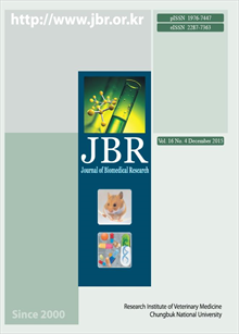
간행물
Journal of Biomedical Research

- 발행기관 충북대학교 동물의학연구소
- 자료유형 학술지
- 간기 계간
- ISSN 1976-7447 (Print)2287-7363 (Online)
- 수록기간 2000 ~ 2015
- 주제분류 의약학 > 수의학 의약학 분류의 다른 간행물
- 십진분류KDC 528DDC 636
권호리스트/논문검색
Vol. 14 No. 1 (2013년 3월) 10건
Review
1.
2013.03
구독 인증기관 무료, 개인회원 유료
Cancer is the result of damage to the genetic system, i.e., dysfunction of the DNA repair system, resulting in dysregulated expression of various molecules, leading to cancer formation, migration, and invasion. In cancer progression, several proteases play a critical role in metastasis; however, their biological mechanism in cancer metastasis is not clearly understood. Among these proteases, cathepsins are a family of lysosomal proteases found in most animal cells. Cathepsins have an important role in protein turnover of mammalian, and are classified into 15 types based on their structure as serine (cathepsin A and G), aspartic (cathepsin D and E), and cysteine cathepsins (cathepsin B, C, F, H, K, L, O, S, V, X, and W). Cysteine cathepsins appear to accelerate the progression of human and rodent cancers, which can be a biomarker of the potency of malignancy or metastasis in mammalian. Overexpression of cyteine cathepsins causes the activation of angiogenesis promoting factor, whereas their downregulation reduces the angiogenesis of cancer progression. Under physiological conditions, cysteine cathepsins are essential in inflammation, infection, and cancer development. Activity of cysteine proteases, i.e., cathepsin B, is required for cancer progression or metastasis. Elevation of cysteine cathepsin is associated with cancer metastasis, angiogenesis, and immunity. Therefore, in this review, we suggest that cysteine cathepsin may be an anticancer target of strong clinical interest, although the exact mechanism of cathepsins in cancer metastasis is under investigation.
4,000원
Original Article
2.
2013.03
구독 인증기관 무료, 개인회원 유료
Amoxicillin, a well-known antibiotic, has a broad spectrum against gram-negative and gram-positive bacteria. This experiment was conducted in order to investigate the effect of micronized and non-micronized amoxicillin prepared using different comminution techniques on change in blood concentration of rats. Forty adult male Sprague Dawley rats (6~7 weeks of age, body weight 128.3 ± 10.7 g) were randomly allocated to two treatment groups: micronized amoxicillin (MA) group treated with micronized amoxicillin trihydrate powder (particle size, over 90% of 10 μm), non-micronized amoxicillin (NMA) group treated with non-micronized amoxicillin trihydrate powder (particle size, over 70% of 100 μm), given 480 mg/kg body weight once daily for four days. The results showed a significant increase in serum concentration in the MA group on days 3 and 4, compared to the NMA group (P<0.05). In particular, serum concentration of the MA group on day 4 was increased almost two times that of the NMA group. The results indicate that due to the increase of the drug’s oral bioavailability, higher serum concentration would be achieved with the micronized amoxicillin trihydrate than with the non-micronized drug.
4,000원
3.
2013.03
구독 인증기관 무료, 개인회원 유료
Taste receptors of the anterior tongue are innervated by the chorda tympani (CT) branch of the facial (VIIth) nerve. The CT nerve transmits information on taste to the ipsilateral nucleus of the solitary tract (NST), which is the first taste central nucleus in the medulla. Taste information is known to be transferred ipsilaterally along the taste pathway in the central nervous system. Some patients with unilateral CT damage often retain their ability to sense taste. This phenomenon is not explained by the unilateral taste pathway. We examined whether neurons in the NST receive information on taste from the contralateral side of the tongue by measuring c-Fos-like Immunoreactivity (cFLI) following taste stimulation of the contralateral side of the tongue in the anesthetized rats. We used four basic taste stimuli, 1.0 M sucrose, 0.3 M NaCl, 0.01 M citric acid, 0.03 M QHCl, and distilled water. Stimulation of one side of the tongue with taste stimuli induced cFLI in the NST bilaterally. The mean number of cFLI ranged from 23.28 ± 2.46 by contralateral QHCl to 30.28 ± 2.26 by ipsilateral NaCl stimulation. The difference between the number of cFLI in the ipsilaterl and contralateral NST was not significant. The result of the current study suggests that neurons in the NST receive information on taste not only from the ipsilateral but also the contralateral side of the tongue.
4,000원
4.
2013.03
구독 인증기관 무료, 개인회원 유료
The objective of this experiment was to assess the relationship between electrical resistance of the vaginal mucosa and plasma progesterone for optimal mating time in the bitch. Eight mature beagle bitches were examined, and we observed eight times of estrus. Vaginal electric resistance was recorded weekly using a Draminski ovulation detector in anestrus, and daily in estrus. Plasma progesterone concentration was estimated by radioimmunoassay. In the bitch, incline in vaginal electric resistance (376.20 ± 105.63 units) showed a closely association with the onset of proestrus. Ovulation day was determined as the first day when plasma progesterone concentration increased above 5.0 ng/ml (Day 0). On Day 0, vaginal mucous electric resistance was 438 ± 132 units. Vaginal mucous electric resistance showed a slight decrease or was maintained until Day 0. However, it showed an explosive increase, and peaked on Day 1~3, which was above 600 units. Two of eight cases peaked on Day 1, three of eight cases were revealed on Day 2, and others were revealed on Day 3. After Day 4, resistance showed a rapid drop to below 600 units and reached 200 units on Day 8. The optimal mating time was determined when vaginal mucous electric resistance was above 600 units.
4,000원
5.
2013.03
구독 인증기관 무료, 개인회원 유료
The aim of this study was to evaluate immunopotentiating activities of β-glucan derived from Saccharomyces (S.) cerevisiae and to select new strains having possibility as an immune-enhancing substance. We examined SB20 strains derived from commercial product as a control, and extracted β-glucans from the four strains of S. cerevisiae. RAW264.7 macrophages were treated with heat-killed yeasts, β-glucans, and lipopolysaccharide (LPS). The production of nitric oxide (NO) and cytokines such as TNF-α and IL-1β were then quantified. When macrophages were induced directly by in vitro addition of β-glucan, little production of NO and IL-1β was observed. When pretreated with strong stimulants, i.e., LPS, most yeasts showed down-modulation of NO and IL-1β production. However, TNF-α secretion was triggered by β-glucans and even more increased by the mixture effect of LPS and β-glucans. In particular, S6 strain induced TNF-α secretion more than other strains. Therefore, we can conclude that the S6 strain has possibility as an immune-enhancing substance.
4,000원
6.
2013.03
구독 인증기관 무료, 개인회원 유료
In this study, we observed anti-diabetic effects of acid hydrolyzed silk peptides, where the amount of peptides in the total amino acid mixture was strictly regulated. Using in vitro diabetes models, silk peptide-containing amino acid mixtures of 5.60% (G5), 11.30% (G10), 14.50% (G15), and 20.50% (G20) were examined separately in order to determine whether they have biological activities. According to our results, a cytoprotective effect was observed following treatment of interleukin-1β in RINm5f pancreas β-cells. As a consequence, Bax, a pro-apoptotic gene, was down-regulated, while Bcl-2, a pro-survival gene, was retained at normal level. Results of the 4’,6-diamidino-2-phentylindole (DAPI) staining assay confirmed that G20 has a better cytoprotective effect. Insulin release from RINm5f cells showed a significant increase following treatment with G5-G20, suggesting that silk peptide effectively regulated and induced insulin production. Single treatment with G5-G20 resulted in enhanced glucose uptake in L6 skeletal muscle cells. In addition, a higher amount of each group inhibited the activity of α-glucosidase. In summary, these data suggest that silk peptide may have an anti-diabetic effect through protection of pancreas β-cells and enhancement of insulin release, which showed a close association with Type 1 diabetes mellitus (DM), and can improve glucose uptake, which was the major target for therapy of Type 2 diabetes. Taken together, we concluded that acid hydrolyzed silk peptides can be used effectively for control of blood sugar metabolism via improvement of the problematic indices of Type 1 and Type 2 DM.
4,000원
7.
2013.03
구독 인증기관 무료, 개인회원 유료
Seong Soo Kang, Jae Kyong Kim, Se Eun Kim, Chun-Sik Bae, Kyung Mi Shim, Seok Hwa Choi, Soon-Jeong Jeong
This study was conducted in order to examine the effects of alcohol-free cetylpyridinium chloride drinking water additive and oral gel on clinical parameters related to periodontal disease in beagle dogs. This study was conducted with healthy 15 beagle dogs. Following a professional teeth cleaning procedure, dogs were divided into three groups. Dogs in the control group received nothing, those in the drinking water additive (DWA) group received 800 ml water with 15 ml of alcohol-free cetylpyridinium chloride drinking water additive daily, and those in the Oral gel (OG) group were treated with oral gel containing alcohol-free cetylpyridinium chloride and 0.05% chlorhexidine gluconate daily. Clinical parameters, including plaque index (PI), calculus index (CI), and gingivitis index (GI) were evaluated at two and four weeks. Dogs in the DWA and OG groups had significantly less plaque than dogs in the control group at two and four weeks (P<0.01, P<0.05). And, at four weeks, CI was significantly lower in the OG group compared to the control group (P<0.05). On GI, similar scores were recorded for all groups during the experimental period. No significant difference was observed between the DWA group and the OG group. The effect of alcohol-free cetylpyridinium chloride drinking water additive was similar to the result for alcohol containing cetylpyridinium chloride mouthwash reported in a previous study. The effect in control of periodontal disease was better in the OG group because of additional chlorhexidine gluconate. However, use of drinking water additive will be more convenient for owners; thus, it will be more effective for achievement of long-term results.
4,000원
8.
2013.03
구독 인증기관 무료, 개인회원 유료
Okjin Kim, Min-Woo Son, Dae-In Moon, Eun-Hye Shin, Hong-Geun Oh, Sunhwa Hong, Sang-Jun Han, Hyun-A Lee, Yung-Ho Chung
The anti-diabetes mechanism of silkworm Bombyx mori L. powder and extracts was found to inhibit the activity of α-glycosidase. The major functional component of silkworm powder was 1-deoxynojirimycin (1-DNJ), which exerts a blood glucose-lowering effect. In this study, we aimed to compare the effects of the supplements, including red ginseng extract on the functional components of silkworm. Fifty silkworm larvae were divided into the control group (Con, N=50), group A (A, artificial diet 95% and mulberry leaf powder 5%), group B (B, artificial diet 95% and mulberry powder 5%), group C (C, artificial diet 95% and Rubus coreanus remainders 5%), group D (D, artificial diet 95% and red ginseng extract 5%), and group E (E, artificial diet 95% and yeast powder (Saccharomyces cerevisiae). Body weights and length of silkworm larvae showed significant improvement in group A, D. In particular, the growth rate in group D (artificial diet 95% and red ginseng extract 5%) was larger than that of Con. In addition, the results showed that 1-DNJ concentration was significantly largest in group D. From these results, it is concluded that the addition of red ginseng extract may be effective for larval growth and 1-DNJ accumulation in silkworm rearing with an artificial diet.
4,000원
Case Report
9.
2013.03
구독 인증기관 무료, 개인회원 유료
Kyu-Shik Jeong, HaiJie Yang, Sun-Hee Do, Eun-Mi Lee, Ah-Young Kim, Eun-Joo Lee, Chang-Woo Min, Kyung-Ku Kang, Myeong-Mi Lee
A 14-year-old female South American sea lion (Otaria byronia) with persistent vaginal secretion and chronic hemorrhagic diarrhea was encountered. During postmortem examination, the uterus was found to resemble a balloon with mucosal congestion and was filled with grayish milky material. The ovaries also had abnormal features, including necrotic surface lesions and multiple whitish foci in the cut section. Hemorrhages and ulcerated changes due to toxemia were observed in other organs, including the liver, spleen, lung, intestines, and lymph nodes. Microscopically, the left ovary contained interlacing fascicles of fibroblast-like cells with blunt-end nuclei showing cytoplasmic positive immunoreactivity against alpha-smooth muscle actin and desmin. The right ovary contained cells with round to cigar-shaped nuclei showing cytoplasmic positive immunoreactivity against vimentin. In conclusion, based on classification of bilateral ovarian tumors as a leiomyoma in the left region and a fibroma in the right region, this sea lion was diagnosed with chronic closed pyometra.
3,000원
10.
2013.03
구독 인증기관 무료, 개인회원 유료
A 10-year-old, castrated male, English cocker spaniel dog was presented for evaluation of a mass in the left forelimb. Physical examination revealed a solitary subcutaneous mass measuring 2.7 × 2.1 × 1 cm in size. Radiographs and ultrasonography showed a well-circumscribed, focally mineralized, non-invasive to muscle layer mass without signs of further bone invasion and periosteal reaction. Cytologic evaluation of the mass through fine needle aspiration revealed a mesenchymal cell type malignant tumor without distant metastasis. An excisional biopsy was performed for definitive diagnosis and the mass was diagnosed as cutaneous hemangiopericytoma. This case report presents disagreement between fine needle aspiration and histopathology during diagnostic procedures of cutaneous hemangiopericytoma in a dog.
3,000원

