간행물
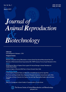
한국동물생명공학회지 (구 한국수정란이식학회지) KCI 등재 Journal of Animal Reproduciton and Biotechnology

- 발행기관 한국동물생명공학회(구 한국수정란이식학회)
- 자료유형 학술지
- 간기 계간
- ISSN 2671-4639 (Print)2671-4663 (Online)
- 수록기간 1983 ~ 2025
- 주제분류 자연과학 > 생물공학 자연과학 분류의 다른 간행물
- 십진분류KDC 527DDC 636
권호리스트/논문검색
Vol.39 No.2 (2024년 6월) 10건
1.
2024.06
구독 인증기관 무료, 개인회원 유료
Background: Post-ovulatory aging (POA) of oocytes is related to a decrease in the quality and quantity of oocytes caused by aging. Previous studies on the characteristics of POA have investigated injury to early embryonic developmental ability, but no information is available on its effects on mitochondrial fission and mitophagy-related responses. In this study, we aimed to elucidate the molecular mechanisms underlying mitochondrial fission and mitophagy in in vitro maturation (IVM) oocytes and a POA model based on RNA sequencing analysis. Methods: The POA model was obtained through an additional 24 h culture following the IVM of matured oocytes. NMN treatment was administered at a concentration of 25 μM during the oocyte culture process. We conducted MitoTracker staining and Western blot experiments to confirm changes in mitochondrial function between the IVM and POA groups. Additionally, comparative transcriptome analysis was performed to identify differentially expressed genes and associated changes in mitochondrial dynamics between porcine IVM and POA model oocytes. Results: In total, 32 common genes of apoptosis and 42 mitochondrial fission and function uniquely expressed genes were detected (≥ 1.5-fold change) in POA and porcine metaphase II oocytes, respectively. Functional analyses of mitochondrial fission, oxidative stress, mitophagy, autophagy, and cellular apoptosis were observed as the major changes in regulated biological processes for oocyte quality and maturation ability compared with the POA model. Additionally, we revealed that the activation of NAD+ by nicotinamide mononucleotide not only partly improved oocyte quality but also mitochondrial fission and mitophagy activation in the POA porcine model. Conclusions: In summary, our data indicate that mitochondrial fission and function play roles in controlling oxidative stress, mitophagy, and apoptosis during maturation in POA porcine oocytes. Additionally, we found that NAD+ biosynthesis is an important pathway that mediates the effects of DRP1-derived mitochondrial morphology, dynamic balance, and mitophagy in the POA model.
4,600원
2.
2024.06
구독 인증기관 무료, 개인회원 유료
Background: Platelet-derived growth factor receptor alpha (PDGFRα) is essential for various biological processes, including fetal Leydig cell differentiation. The PDGFRαEGFP mouse model, which expresses an eGFP fusion gene under the native Pdgfrα promoter, serves as a valuable resource for exploring PDGFRα’s expression and function in vivo. This study investigates PDGFRα expression in adult testicular cells using PDGFRαEGFP mouse model. Methods: Genotyping PCR and gel electrophoresis were used to confirm the zygosity of PDGFRαEGFP mice. Histological examination and fluorescence imaging were used to identify PDGFRα expression within testicular tissue. Immunohistochemical analysis assessed the co-expression of PDGFRα with c-Kit, ANO-1, and TASK-1 in testicular cells. Results: Genotyping confirmed the heterozygous status of the mice, which is crucial for studies due to the embryonic lethal phenotype observed in homozygotes. Histological and fluorescence imaging revealed that PDGFRα+ cells were primarily located in the interstitial spaces of the testis, specifically within Leydig cells and peritubular myoid cells (PMCs). Immunohistochemical results showed PDGFRα co-localization with c-Kit and ANO-1 in Leydig cells and a complete co-localization with TASK-1 in both Leydig cells and PMCs. Conclusions: The findings demonstrate specific expression of PDGFRα in Leydig cells and PMCs in adult testicular tissue. The co-expression of PDGFRα with c-Kit, ANO-1, and TASK-1 suggests complex regulatory mechanisms, possibly influencing testicular function and broader physiological processes.
4,000원
3.
2024.06
구독 인증기관 무료, 개인회원 유료
Background: Using cryovial for freezing dog spermatozoa provides a practical method to increase extended sperm volume and shorten the time required for equilibration by using a simple freezing techniques. The purpose of this study was to determine the optimal thawing condition for dog sperm cryopreservation using cryovials. Methods: For sperm freezing, cryovials with 200 × 106 sperm/mL were cooled after the addition of tris egg yolk extender (TEY) at 4℃ for 20 min, then TEY with 4% glycerol was added and equilibrated for another 20 min before being aligned over LN2 vapor for another 20 min and plunged directly into LN2. Spermatozoa were thawed in a water bath at 37℃ for varying times (25 sec, 60 sec, 90 sec, and 120 sec) in the first experiment. In the second experiment, spermatozoa were thawed in a water bath at various temperatures and times (37℃ for 1 min, 37℃ for 1 min with gentle stirring, 24℃ for 24 min, and 75℃ for 20 sec). In these experiments, the effect of thawing conditions on motility parameters, viability (SYBR-14/PI), and acrosome integrity (PSA/ FITC) of spermatozoa were investigated. Results: The post-thaw sperm motility parameters, viability, and acrosome integrity were not significantly different across the experimental groups. Conclusions: In this study, the characteristics of spermatozoa frozen using cryovials were not significantly affected by various thawing conditions.
4,000원
4.
2024.06
구독 인증기관 무료, 개인회원 유료
Background: In healthy dentin conditions, odontoblasts have an important role such as protection from invasion of pathogens. In mammalian teeth, progenitors such as mesenchymal stem cells (MSCs) can migrate and differentiate into odontoblast-like cells, leading to the formation of reparative dentin. For differentiation using stem cells, it is crucial to provide conditions similar to the complex and intricate in vivo environment. The purpose of this study was to evaluate the potential of differentiation into odonto/ osteoblasts, and compare co-culture with/without epithelial cells. Methods: MSCs and epithelial cells were successfully isolated from dental tissues. We investigated the influences of epithelial cells on the differentiation process of dental pulp stem cells into odonto/osteoblasts using co-culture systems. The differentiation potential with/without epithelial cells was analyzed for the expression of specific markers and calcium contents. Results: Differentiated odonto/osteoblast derived from dental pulp tissue-derived mesenchymal stem cells with/without epithelial cells were evaluated by qRT-PCR, immunostaining, calcium content, and ALP staining. The expression of odonto/ osteoblast-specific markers, calcium content, and ALP staining intensity were significantly increased in differentiated cells. Moreover, the odonto/osteogenic differentiation capacity with epithelial cells co-culture was significantly higher than without epithelial cells co-culture. Conclusions: These results suggest that odonto/osteogenic differentiation co-cultured with epithelial cells has a more efficient application.
4,000원
5.
2024.06
구독 인증기관 무료, 개인회원 유료
Background: Aflatoxin B1 (AFB1) is a toxic metabolite generated by Aspergillus species and is commonly detected during the processing and storage of food; it is considered a group I carcinogen. The hepatotoxic effects, diseases, and mechanisms induced by AFB1 owing to chronic or acute exposure are well documented; however, there is a lack of research on its effects on the intestine, which is a crucial organ in the digestive process. Dogs are often susceptible to chronic AFB1 exposure owing to lack of variation in their diet, unlike humans, thereby rendering them prone to its effects. Therefore, we investigated the effects of AFB1 on canine small intestinal epithelial primary cells (CSIc). Methods: We treated CSIc with various concentrations of AFB1 (0, 1.25, 2.5, 5, 10, 20, 40, and 80 μM) for 24 h and analyzed cell viability and transepithelial-transendothelial electrical resistance (TEER) value. Additionally, we analyzed the mRNA expression of tight junction-related genes (OCLN, CLDN3, TJP1, and MUC2), antioxidant-related genes (CAT and GPX1), and apoptosis-related genes (BCL2, Bax, and TP53). Results: We found a significant decrease in CSIc viability and TEER values after treatment with AFB1 at concentrations of 20 μM or higher. Quantitative polymerase chain reaction analysis indicated a downregulation of OCLN, CLDN3, and TJP1 in CSIc treated with 20 μM or higher concentrations of AFB1. Additionally, AFB1 treatment downregulated CAT , GPX1, and BCL2. Conclusions: Acute exposure of CSIc to AFB1 induces toxicity, and exposure to AFB1 above a certain threshold compromises the barrier integrity of CSIc.
4,000원
6.
2024.06
구독 인증기관 무료, 개인회원 유료
Background: The Hanwoo industry must develop technologies that can increase the production of preferred cuts to match changing consumer trends. In this study, we aimed to estimate the genetic parameters for carcass traits (carcass weight, eye muscle area, back fat thickness, and intramuscular fat) and primal cut traits (tenderloin, loin, strip loin, neck, clod, top round, bottom round, brisket, shank, and rib) in a Hanwoo population to obtain basic data for improving primal cut productivity. Methods: Data from 1,905 Hanwoo steers, including carcass traits and primal cut weights, were collected. Genetic parameters were estimated using REMLF90 in a multi-trait analysis. Results: High heritability was found for carcass weight (0.52) and strip loin yield (0.63). Genetic correlations between carcass weight and primal cut weights ranged from 0.52 to 0.93. Conclusions: This study demonstrates the significant potential for genetic improvement in Hanwoo cattle through selective breeding, particularly for traits with high heritability and genetic correlations. These findings provide crucial insights into optimizing breeding programs to improve Hanwoo cattle production efficiency.
4,000원
7.
2024.06
구독 인증기관 무료, 개인회원 유료
Background: This study focused on reproductive traits in Hanwoo cattle, specifically the environmental factors affecting gestation length and birth weight. Methods: The records of 1,540 cows calved at the Hanwoo Research Institute from 2015 to 2023 were examined. This study analyzed two populations, linebreeding Hanwoo (LBH) and general Hanwoo (GH), with all cows undergoing estrus synchronization and artificial insemination. The R software was used to compare the differences between the two populations and analyze the environmental factors affecting each trait. Results: The results showed that the average gestation length for LBH was 283.28 ± 5.93 days, which was significantly shorter than that of the GH, which had an average of 285.63 ± 6.21 days (p < 0.001). The average birth weight of LBH calves was 25.10 ± 3.69 kg, significantly lighter than GH calves, which weighed 27.26 ± 4.11 kg on average (p < 0.001). Analysis of environmental factors revealed significant differences in the gestation length of LBH based on dam parity, year, and season of calving. However, no significant differences were observed based on calf sex. For LBH, birth weight showed significant differences based on dam parity, year of calving, and sex of the calf, but not the season of calving. In GH, gestation length varied with dam parity and calving season, but not with calving year or calf sex. The GH birth weight showed differences based on dam parity, year of calving, and calf sex, but not the season of calving. Conclusions: Reproductive traits in the Hanwoo cattle industry are economically vital but are heavily influenced by environmental factors due to their low heritability. An accurate evaluation of the genetic potential of these traits requires an analysis of the environmental factors affecting them. The results of this study serve as foundational data for predicting the potential for genetic improvement in the gestation length and birth weight of Hanwoo cattle.
4,000원
8.
2024.06
구독 인증기관 무료, 개인회원 유료
Shuntaro Miura, Heejae Kang, Seonggyu Bang, Ayeong Han, Islam M. Saadeldin, Sanghoon Lee, Koichi Takimoto, Jongki Cho
Background: Porcine embryonic development is widely utilized in the medical industry. However, the blastocyst development rate in vitro is lower compared to in vivo . To address this issue, various supplements are employed. Extracellular vesicles (EVs) play the role of communicators that carry many bioactive cargoes. Additionally, the contents of EVs can vary on the estrous cycle. Methods: We compared the effects of adding EVs derived from porcine uterine fluid (UF), categorized as non-EV (G1), EVs in estrus (G2) and EVs in diestrus (G3). After in vitro culture (IVC) was performed in three different groups, cleavage rate and blastocyst development rate were examined. In addition, glutathione (GSH) and reactive oxygen species (ROS) levels were measured 2 days after activation to assess oxidative stress. Results: Using NTA and cryo-TEM, we confirmed the presence of EVs with sizes ranging from 30 nm to 200 nm, that the particles were suitable for analysis for analysis. In IVC data, the highest cleavage rate was observed in G2, which was significantly different from G1 but not significantly different from the next highest, G3. Similarly, the highest blastocyst development rate was observed in G2, which was significantly different from G1 but not significantly different from the next highest, G3. Conclusions: These results indicate that estrus derived EVs contain biofactors beneficial for early blastocyst development, including GSH which protects the blastocyst from oxidative stress. Additionally, although diestrus-derived EVs are expected to have some effect on blastocyst development, it appeared to be less effective than estrus-derived EVs.
4,000원
9.
2024.06
구독 인증기관 무료, 개인회원 유료
Background: As the number of households raising companion dogs increases, the pet genetic analysis market also continues to grow. However, most studies have focused on specific purposes or native breeds. This study aimed to collect genomic data through single nucleotide polymorphism (SNP) chip analysis of companion dogs in South Korea and perform genetic diversity analysis and SNP annotation. Methods: We collected samples from 95 dogs belonging to 26 breeds, including mixed breeds, in South Korea. The SNP genotypes were obtained for each sample using an Axiom™ Canine HD Array. Quality control (QC) was performed to enhance the accuracy of the analysis. A genetic diversity analysis was performed for each SNP. Results: QC initially selected SNPs, and after excluding non-diverse ones, 621,672 SNPs were identified. Genetic diversity analysis revealed minor allele frequencies, polymorphism information content, expected heterozygosity, and observed heterozygosity values of 0.220, 0.244, 0.301, and 0.261, respectively. The SNP annotation indicated that most variations had an uncertain or minimal impact on gene function. However, approximately 16,000 non-synonymous SNPs (nsSNPs) have been found to significantly alter gene function or affect exons by changing translated amino acids. Conclusions: This study obtained data on SNP genetic diversity and functional SNPs in companion dogs raised in South Korea. The results suggest that establishing an SNP set for individual identification could enable a gene-based registration system. Furthermore, identifying and researching nsSNPs related to behavior and diseases could improve dog care and prevent abandonment.
4,000원
10.
2024.06
구독 인증기관 무료, 개인회원 유료
Background: Despite its anticancer activity, cisplatin exhibits severe testicular toxicity when used in chemotherapy. Owing to its wide application in cancer therapy, the reduction of damage to normal tissue is of imminent clinical need. In this study, we evaluated the effects of catechin hydrate, a natural flavon-3-ol phytochemical, on cisplatin-induced testicular injury. Methods: Type 2 mouse spermatogonia (GC-1 spg cells) were treated with 0-100 μM catechin and cisplatin. Cell survival was estimated using a cell proliferation assay and Ki-67 immunostaining. Apoptosis was assessed via flow cytometry with the Dead Cell Apoptosis assay. To determine the antioxidant effects of catechin hydrate, Nrf2 expression was measured using qPCR and CellROX staining. The anti-inflammatory effects were evaluated by analyzing the gene and protein expression levels of iNOS and COX2 using qPCR and immunoblotting. Results: The 100 μM catechin hydrate treatment did not affect healthy GC-1 spg cells but, prevented cisplatin-induced GC-1 spg cell death via the regulation of anti-oxidants and inflammation-related molecules. In addition, the number of apoptotic cells, cleaved-caspase 3 level, and BAX gene expression levels were significantly reduced by catechin hydrate treatment in a cisplatin-induced GC-1 spg cell death model. In addition, antioxidant and anti-inflammatory marker genes, including Nrf2 , iNOS, and COX2 were significantly downregulated by catechin hydrate treatment in cisplatintreated GC-1 cells. Conclusions: Our study contributes to the opportunity to reintroduce cisplatin into systemic anticancer treatment, with reduced testicular toxicity and restored fertility.
4,000원

