간행물
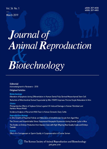
한국동물생명공학회지 (구 한국수정란이식학회지) KCI 등재 Journal of Animal Reproduciton and Biotechnology

- 발행기관 한국동물생명공학회(구 한국수정란이식학회)
- 자료유형 학술지
- 간기 계간
- ISSN 2671-4639 (Print)2671-4663 (Online)
- 수록기간 1983 ~ 2025
- 주제분류 자연과학 > 생물공학 자연과학 분류의 다른 간행물
- 십진분류KDC 527DDC 636
권호리스트/논문검색
Vol. 33 No. 2 (2018년 6월) 5건
Clinics
1.
2018.06
구독 인증기관 무료, 개인회원 유료
Ensuring timely ovulation concerning the service is valuable. A satisfactory conception rate can be achieved by making sure that ovulation occurs within 7-18 hours after artificial insemination (AI). Delayed ovulation is one of the disturbances commonly encountered in repeat breeding animals. Although demanding research, many studies have not been conducted. Therefore, we aimed to examine the relation between ovulation confirmation and conception rate in dairy cattle. The research findings showed that the signs of true estrus were bred 12 hours after the onset of estrus by AI in cattle. Also, the performance of AI on ovulation was confirmed by the presence of fluctuant Graafian follicles through rectal palpation. From the results, we confirmed that cow encountered delayed ovulation were bred again. The Conception rate in cows with confirmed ovulation was 51.9%, while for those without confirmed ovulation were 33.3%. In conclusion, the results indicate that ovulation confirmation will likely increase conception rate.
4,000원
Reproduction
2.
2018.06
구독 인증기관 무료, 개인회원 유료
The major focus of this study is to analyze the expression of bovine MMPs and to monitor their activity during the estrus cycle and pregnancy. During pregnancy, MMP-2 expression was detectable around 30 days but became insignificant by 60 days, then started to increase again around 90 days and reached the maximum at 250 days. The activity of MMP-2 protein changed in accordance with its expression level. As expected, the level of TIMP-2 exhibited a reverse pattern. About MMP-9, high level expression was observed as early as 30 days and gradually increase until 90 days. Then started to decrease after 250 days. Again, the sites of MMP-9 expression were similar to those of MMP-2. On the other hand, expression of TIMP-3 remained low until 90 days but showed a small and temporal increase around 250 days. In summary, expression of different MMPs were differentially regulated during estrus cycle and pregnancy. While the expression of MMP-2 was high in estrus cycle, MMP-9 slowly takes over with the progression of pregnancy. These results indicated that the luteal tissue perform distinct functions during pregnancy and estrus. Perhaps the activity of MMP-2 is required for the structural remodeling of luteum, resulting the suppression of P4 inflow from blood. On the other hand, steady maintenance of MMP-9 throughout luteal development is important for the activation of cell proliferation, maturation and angiogenesis.
4,000원
3.
2018.06
구독 인증기관 무료, 개인회원 유료
This study was conducted to examine the influences of two human chorion gonadotrophins (hCGs) being injected into young or aged (45- to 65-week old) outbred (ICR) mice on developmental capacity of oocytes retrieved. In vitro-culture and parthenogenetic activation of oocytes retrieved were employed for the assessment. Superovulation was determined as being induced when more than 25 oocytes were retrieved. No aged mice were superovulated, while in contrast, 67-100% were superovulated in the 6- to 8-week-old (young) mice. In the aged, hCG injection yielded better retrieval (5 vs. 13 to 14.8 oocytes/mouse). Overall, no significant difference between two hCGs was detected but between the young and aged, significant differences in maturational arrest (0% vs. 39% MI arrest and 46% vs. 15% degeneration) and developmental capacity (24% vs. 46% 8-cell embryo development) were detected. In conclusion, hCG injection contributes to increasing oocyte retrieval from aged outbred mice, but the kinds of gonadotrophin influenced the efficiency of hyperstimulation induction in specific ages.
4,000원
Semen
4.
2018.06
구독 인증기관 무료, 개인회원 유료
Establishment of Normal Reference Data of Analysis in the Fresh and Cryopreserved Canine Spermatozoa
The cryopreservation has been extensively applied in many cells including spermatozoa (semen) during past several decades. Especially, the canine spermatozoa cryopreservation has contributed on generation of progeny of rare/genetically valuable dog breeds, genome resource banking and transportation of male germplasm at a distant place. However, severe and irreversible damages to the spermatozoa during cryopreservation procedures such as the thermal shock (cold shock), formation of intracellular ice crystals, osmotic shock, stress of cryoprotectants and generator of reactive oxygen species (ROS) have been addressed. According as a number of researches have been conducted to overcome these problems and to advance cryopreservation technique, several analytical methods have been employed to evaluate the quality of the fresh or cryopreserved canine spermatozoa in regards to the motility, morphology, integrity of membrane and DNA, mitochondrial activity, ROS generation, binding affinity to oocytes, in vitro fertilization potential and fertility potential by artificial insemination. Because the study designs with certain application of analytical methods are selective and varied depending on each experimental objective and laboratory condition, it is necessary to establish the normal reference data of the fresh or cryopreserved canine spermatozoa for each analytical method to monitor experimental procedure, to translate raw data and to discuss results. Here, we reviewed the recent articles to introduce various analytical methods for the canine spermatozoa as well as to establish the normal reference data for each analytical method in the fresh or cryopreserved canine spermatozoa, based on the results of the previous articles. We hope that this review contributes to the advancement of cryobiology in canine spermatozoa.
4,000원
Stem Cells
5.
2018.06
구독 인증기관 무료, 개인회원 유료
The trans-differentiation potential of mesenchymal stem cells (MSCs) is employed, but there is little understanding of the cell source-dependent trans-differentiation potential of MSCs into corneal epithelial cells. In the present study, we induced trans-differentiation of MSCs derived from umbilical cord matrix (UCM-MSCs) and from dental tissue (D-MSCs), and we comparatively evaluated the in vitro trans-differentiation properties of both MSCs into corneal epithelial-like cells. Specific cell surface markers of MSC (CD44, CD73, CD90, and CD105) were detected in both UCM-MSCs and D-MSCs, but MHCII and CD119 were significantly lower (P < 0.05) in UCM-MSCs than in D-MSCs. In UCM-MSCs, not only expression levels of Oct3/4 and Nanog but also proliferation ability were significantly higher (P < 0.05) than in D-MSCs. In vitro differentiation abilities into adipocytes and osteocytes were confirmed for both MSCs. UCM-MSCs and D-MSCs were successfully trans-differentiated into corneal epithelial cells, and expression of lineage-specific markers (Cytokeratin-3, -8, and -12) were confirmed in both MSCs using immunofluorescence staining and qRT-PCR analysis. In particular, the differentiation capacity of UCM-MSCs into corneal epithelial cells was significantly higher (P < 0.05) than that of D-MSCs. In conclusion, UCM-MSCs have higher differentiation potential into corneal epithelial-like cells and have lower expression of CD119 and MHC class II than D-MSCs, which makes them a better source for the treatment of corneal opacity.
4,500원

