간행물
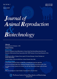
한국동물생명공학회지 (구 한국수정란이식학회지) KCI 등재 Journal of Animal Reproduciton and Biotechnology

- 발행기관 한국동물생명공학회(구 한국수정란이식학회)
- 자료유형 학술지
- 간기 계간
- ISSN 2671-4639 (Print)2671-4663 (Online)
- 수록기간 1983 ~ 2025
- 주제분류 자연과학 > 생물공학 자연과학 분류의 다른 간행물
- 십진분류KDC 527DDC 636
권호리스트/논문검색
Vol. 37 No. 2 (2022년 6월) 11건
1.
2022.06
구독 인증기관 무료, 개인회원 유료
Currently, there is no treatment to reverse or cure heart failure caused by ischemic heart disease and myocardial infarction despite the remarkable advances in modern medicine. In addition, there is a lack of evidence regarding the existence of stem cells involved in the proliferation and regeneration of cardiomyocytes in adult hearts. As an alternative solution to overcome this problem, protocols for differentiating human pluripotent stem cell (hPSC) into cardiomyocyte have been established, which further led to the development of cell therapy in major leading countries in this field. Recently, clinical studies have confirmed the safety of hPSC-derived cardiac progenitor cells (CPCs). Although several institutions have shown progress in their research on cell therapy using hPSC-derived cardiomyocytes, the functions of cardiomyocytes used for transplantation remain to be those of immature cardiomyocytes, which poses a risk of graft-induced arrhythmias in the early stage of transplantation. Over the last decade, research aimed at achieving maturation of immature cardiomyocytes, showing same characteristics as those of mature cardiomyocytes, has been actively conducted using various approaches at leading research institutes worldwide. However, challenges remain in technological development for effective generation of mature cardiomyocytes with the same properties as those present in the adult hearts. Therefore, in this review, we provide an overview of the technological development status for maturation methods of hPSC-derived cardiomyocytes and present a direction for future development of maturation techniques.
4,500원
2.
2022.06
구독 인증기관 무료, 개인회원 유료
The ability to determine the sex of bovine embryos before the transfer is advantageous in livestock management, especially in dairy production, where female calves are preferred in milk industry. The milk production of female and male cattle benefits both the dairy and beef industries. Pre-implantation sexing of embryos also helps with embryo transfer success. There are two approaches for sexing bovine embryos in farm animals: invasive and non-invasive. A non-invasive method of embryo sexing retains the embryo’s autonomy and, as a result, is less likely to impair the embryo’s ability to move and implant successfully. There are lists of non-invasive embryo sexing such as; Detection of H-Y antigens, X-linked enzymes, and sexing based on embryo cleavage and development. Since it protects the embryo’s autonomy, the non-invasive procedure is considered to be the safest. Invasive methods affect an embryo’s integrity and are likely to damage the embryo’s chances of successful transformation. There are different types of invasive methods such as polymerase chain reaction, detection of male chromatin Y chromosomespecific DNA probes, Loop-mediated isothermal amplification (LAMP), cytological karyotyping, and immunofluorescence (FISH). The PCR approach is highly sensitive, precise, and effective as compared to invasive methods of farm animal embryonic sexing. Invasive procedures, such as cytological karyotyping, have high accuracy but are impractical in the field due to embryonic effectiveness concerns. This technology can be applicable especially in the dairy and beef industry by producing female and male animals respectively. Enhancing selection accuracy and decreasing the multiple ovulation embryo transfer costs.
4,000원
3.
2022.06
구독 인증기관 무료, 개인회원 유료
Receptor tyrosine kinase c-Kit, a marker found on interstitial cells of Cajal (ICCs), is expressed in Leydig cells, which are testicular interstitial cells. The expression of other ICC markers has not yet been reported. In this study, we investigated the expression of c-Kit and anoctamin 1 (ANO1), another ICC marker, in mouse testes. In addition, the relationship between c-Kit and ANO1 expression and Leydig cell function was investigated. We observed that c-Kit and ANO1 were predominantly expressed in mouse Leydig cells. The mRNA and protein of c-Kit and ANO1 were expressed in TM3, a mouse Leydig cell line. LH induced an increase in intracellular Ca2+ concentration, membrane depolarization, and testosterone secretion, whereas these signals were inhibited in the presence of c-Kit and ANO1 inhibitors. These results show that c-Kit and ANO1 are expressed in Leydig cells and are involved in testosterone secretion. Our findings suggest that Leydig cells may act as ICCs in testosterone secretion.
4,000원
4.
2022.06
구독 인증기관 무료, 개인회원 유료
Hwan-Deuk Kim, Hye-Jin Jeon, Min Jang, Seul-Gi Bae, Sung-Ho Yun, Jee-Eun Han, Seung-Joon Kim, Won-Jae Lee
The ovary undergoes substantial physiological changes along with estrus phase to mediate negative/positive feedback to the upstream reproductive tissues and to play a role in producing a fertilizable oocyte in the developing follicles. However, the disorder of estrus cycle in female can lead to diseases, such as cystic ovary which is directly associated with decline of overall reproductive performance. In gene expression studies of ovaries, quantitative reverse transcription polymerase chain reaction (qPCR) assay has been widely applied. During this assay, although normalization of target genes against reference genes (RGs) has been indispensably conducted, the expression of RGs is also variable in each experimental condition which can result in false conclusion. Because the understanding for stable RG in porcine ovaries was still limited, we attempted to assess the stability of RGs from the pool of ten commonly used RGs (18S, B2M, PPIA, RPL4, SDHA, ACTB, GAPDH, HPRT1, YWHAZ, and TBP) in the porcine ovaries under different estrus phase (follicular and luteal phase) and cystic condition, using stable RG-finding programs (geNorm, Normfinder, and BestKeeper). The significant (p < 0.01) differences in Ct values of RGs in the porcine ovaries under different conditions were identified. In assessing the stability of RGs, three programs comprehensively agreed that TBP and YWHAZ were suitable RGs to study porcine ovaries under different conditions but ACTB and GAPDH were inappropriate RGs in this experimental condition. We hope that these results contribute to plan the experiment design in the field of reproductive physiology in pigs as reference data.
4,000원
5.
2022.06
구독 인증기관 무료, 개인회원 유료
Cryopreservation of porcine ovarian tissue by vitrification method is a promising approach to preserve genetic materials for future use. However, information is not enough and technology still remains in a challenge stage in pig. Therefore, the objective of present study was to determine possibility of vitrification method to cryopreserve porcine ovarian tissue and to confirm an occurrence of cryoinjuries. Briefly, cryoinjuries and apoptosis patterns in vitrified-warmed ovarian tissue were examined by histological evaluation and TUNEL assay respectively. In results, a damaged morphology of oocytes was detected among groups and the rate was significantly (p < 0.05) lower in vitrification group (25.8%) than freezing control group (67.7%), while fresh control group (6.6%) showed significantly (p < 0.05) lower than both groups. In addition, cryoinjury that form a wave pattern of tissues around follicles was found in the frozen control group, but not in the fresh control group as well as in the vitrification group. Apoptotic cells in follicle was observed only in freezing control group while no apoptotic cell was found in both fresh control and vitrification. Similarly, apoptotic patterns of tissues not in follicle were comparable between fresh control and vitrification groups while freezing control group showed increased tendency. Conclusively, it was confirmed that vitrification method has a prevention effect against cryoinjury and this method could be an alternative approach for cryopreservation of genetic material in pigs. Further study is needed to examine the viability of oocytes derived from vitrified-warmed ovarian tissue.
4,000원
6.
2022.06
구독 인증기관 무료, 개인회원 유료
Sperm cryopreservation is a fundamental process for the long-term conservation of livestock genetic resources. Yet, the packaging method has been shown, among other factors, to affect the frozen-thawed (FT) sperm quality. This study aimed to develop a new mini-straw for sperm cryopreservation. In addition, the kinematic patterns, viability, acrosome integrity, and mitochondrial membrane potential (MMP) of boar spermatozoa frozen in the developed 0.25 mL straw, 0.25 mL (minitube, Germany), or 0.5 mL (IMV technologies, France) straws were assessed. Postthaw kinematic parameters were not different (experiment 1: total motility (33.89%, 32.42%), progressive motility (19.13%, 19.09%), curvilinear velocity (42.32, 42.86), and average path velocity (33.40, 33.62) for minitube and the developed straws, respectively. Further, the viability (38.56%, 34.03%), acrosome integrity (53.38%, 48.88%), MMP (42.32%, 36.71%) of spermatozoa frozen using both straw were not differ statistically (p > 0.05). In experiment two, the quality parameters for semen frozen in the developed straw were compared with the 0.5 mL IMV straw. The total motility (41.26%, 39.1%), progressive motility (24.62%, 23.25%), curvilinear velocity (46.44, 48.25), and average path velocity (37.98, 39.12), respectively, for IMV and the developed straw, did not differ statistically. Additionally, there was no significant difference in the viability (39.60%, 33.17%), acrosome integrity (46.23%, 43.23%), and MMP (39.66, 32.51) for IMV and the developed straw, respectively. These results validate the safety and efficiency of the developed straw and highlight its great potential for clinical application. Moreover, both 0.25 mL and 0.5 mL straws fit the present protocol for cryopreservation of boar spermatozoa.
4,000원
7.
2022.06
구독 인증기관 무료, 개인회원 유료
Jeongwoo Kwon, Yu-Jin Jo, Seung-Bin Yoon, Hyeong-ju You, Changsic Youn, Yejin Kim, Jiin Lee, Nam-Hyung Kim, Ji-Su Kim
Heterogeneous nuclear ribonucleoprotein A2/B1 (hnRNPA2/B1) is an N6-methyladenosine (m6A) RNA modification regulator and a key determinant of premRNA processing, mRNA metabolism and transportation in cells. Currently, m6A reader proteins such as hnRNPA2/B1 and YTHDF2 has functional roles in mice embryo. However, the role of hnRNPA2/B1 in porcine embryogenic development are unclear. Here, we investigated the developmental competence and mRNA expression levels in porcine parthenogenetic embryos after hnRNPA2/B1 knock-down. HhnRNPA2/B1 was localized in the nucleus during subsequent embryonic development since zygote stage. After hnRNPA2/B1 knock-down using double stranded RNA injection, blastocyst formation rate decreased than that in the control group. Moreover, hnRNPA2/B1 knock-down embryos show developmental delay after compaction. In blastocyste stage, total cell number was decreased. Interestingly, gene expression patterns revealed that transcription of Pou5f1, Sox2, TRFP2C, Cdx2 and PARD6B decreased without changing the junction protein, ZO1, OCLN, and CDH1. Thus, hnRNPA2/B1 is necessary for porcine early embryo development by regulating gene expression through epigenetic RNA modification.
4,000원
8.
2022.06
구독 인증기관 무료, 개인회원 유료
Epididymal sperm cryopreservation provides a potential method for preserving genetic material from males of endangered species. This pilot study was conducted to develop a freezing method for tiger epididymal sperm. We evaluated post-thaw sperm condition using testes with intact epididymides obtained from a Siberian tiger (Panthera tigris altaica ) after castration. The epididymis was chopped in Tyrode's albumin-lactate-pyruvate 1x and incubated at 5% CO2, 95% air for 10 min. The Percoll separation density gradient method was used for selective recovery of motile spermatozoa after sperm collection using a cell strainer. The spermatozoa were diluted with modified Norwegian extender supplemented with 20 mM trehalose (extender 1) and subsequent extender 2 (extender 1 with 10% glycerol) and frozen using LN2 vapor. After thawing at 37℃ for 25 s, Isolate® solution was used for more effective recovery of live sperm. Sperm motility (computerized assisted sperm analysis, CASA), viability (SYBR-14 and Propidium Iodide) and acrosome integrity (Pisum sativum agglutinin with FITC) were evaluated. The motility of tiger epididymal spermatozoa was 40.1 ± 2.0%, and progressively motile sperm comprised 32.7 ± 2.3%. Viability was 56.3 ± 1.6% and acrosome integrity was 62.3 ± 4.4%. Cryopreservation of tiger epididymal sperm using a modified Norwegian extender and density gradient method could be effective to obtain functional spermatozoa for future assisted reproductive practices in endangered species.
4,000원
9.
2022.06
구독 인증기관 무료, 개인회원 유료
Recent progress has been made to establish intestinal organoids for an in vitro model as a potential alternative to an in vivo system in animals. We previously reported a reliable method for the isolation of intestinal crypts from the small intestine and robust three-dimensional (3D) expansion of intestinal organoids (basal-out) in adult bovines. The present study aimed to establish next-generation intestinal organoids for practical applications in disease modeling-based host-pathogen interactions and feed efficiency measurements. In this study, we developed a rapid and convenient method for the efficient generation of intestinal organoids through the modulation of the Wnt signaling pathway and continuous apical-out intestinal organoids. Remarkably, the intestinal epithelium only takes 3-4 days to undergo CHIR (1 µM) treatment as a Wnt activator, which is much shorter than that required for spontaneous differentiation (7 days). Subsequently, we successfully established an apical-out bovine intestinal organoid culture system through suspension culture without Matrigel matrix, indicating an apical-out membrane on the surface. Collectively, these results demonstrate the efficient generation and next-generation of bovine intestinal organoids and will facilitate their potential use for various purposes, such as disease modeling, in the field of animal biotechnology.
4,000원
10.
2022.06
구독 인증기관 무료, 개인회원 유료
Imperforate hymen is a rare congenital disorder that may predispose to retention of fluid in the vagina and uterus, thereby resulting in conditions such as hematocolpos, pyocolpos, and pyocolpometra in female dogs. A 7-year-old intact female shih tzu exhibiting abdominal distension, depression, anorexia, dysuria, dyschezia, and tenesmus was diagnosed with pyocolpos; a 9-year-old intact female Yorkshire terrier with abdominal mass, dysuria, and tenesmus was diagnosed with hematocolpos; and a 7-year-old intact female shih tzu with dysuria, dyschezia, anorexia, and vomiting was diagnosed with pyocolpometra. Ovariohysterectomy and partial vaginectomy were performed, and the blind end of the vaginal stump was omentalized. This clinical report provide diagnostic process and surgical treatment option for congenital vaginal obstruction cases.
4,000원
11.
2022.06
구독 인증기관 무료, 개인회원 유료
Busulfan is the most commonly used drug for preconditioning during the transplantation of hematopoietic stem cells and male germ cells. Here, we describe side effects of high doses of busulfan in male mongrel dogs. Busulfan was intravenously administered to three groups of dogs at doses of 10, 15, and 17.5 mg/kg body weight. The total white blood cell, neutrophil, eosinophil, lymphocyte, monocyte, and platelet counts steadily reduced in a dose-dependent manner following busulfan treatment. The white blood cell, neutrophil, and monocyte counts recovered after 6 weeks of busulfan treatment, however, the eosinophil, lymphocyte, and platelet counts remained unaltered. Additionally, there was one fatality in the each of the groups that were administered 15 and 17.5 mg/kg busulfan. The gross lesions included severe hemorrhage in the stomach, intestinal tracts, mesentery and urinary bladder. Microscopic investigation revealed severe pulmonary edema and hemorrhage in the lungs, and severe multifocal to coalescing transmural hemorrhage in the intestines and urinary bladder. These results indicated that treatment with busulfan at doses higher than 15 mg/kg initiates severe bleeding in the internal organs and can have fatal results.
4,000원

